Ankle Ultrasound
Disorders around the ankle are common, both as acute injuries and chronic disorders. Various imaging modalities are available to evaluate the ankle, including X-rays, MRI, and ultrasound. Ultrasound has the advantage of providing a dynamic assessment of the ligaments and tendons[1].

Achilles tendon rupture
The Achilles tendon is the strongest tendon in the body. It is in the back of the lower calf and connects the calf muscles to the heel bone (see Figure 1). Acute Achilles tendon tears occur in the setting of sports that involve running and jumping, in particular during phases of sudden increased sports participation. Patients often report the sensation of sudden acute pain and an audible pop, with the feeling of having been kicked in the calf.
Ultrasound can confirm a suspected ruptured Achilles tendon (Figure 2). It can characterise the tear as partial or full thickness and allows measurement of the tear gap in various degrees of ankle flexion. This information will help the orthopaedic surgeon to recommend the best treatment options. The overall sensitivity of ultrasound for detecting complete Achilles tendon ruptures is 94.8% and the specificity is 98.7%[2].

Achilles tendonitis
Achilles tendonitis is an overuse disorder that causes pain at the back of the lower calf and the heel. It is also referred to as Achilles tendonitis, Achilles tendinosis, and Achilles tendinopathy. It is caused by chronic overuse of the tendon, leading to repetitive micro-injuries. Although it more commonly occurs in athletes, volleyball and basketball players, anyone can be affected. Certain conditions of the foot, such as a flat foot deformity, or a more square-shaped morphology of the heel bone, the so-called Haglund’s deformity of the calcaneus, can predispose to Achilles tendinitis. Besides pain, patients often notice swelling or a lump at the back of the heel.
Ultrasound can readily visualise the entire length of the Achilles tendon from the calcaneus to the calf muscles. The diseased Achilles tendon becomes thickened and ‘dark’ on ultrasound with loss of the bright striated pattern characteristic of a healthy tendon (see Figure 3).
Tibialis posterior and peroneal tendinopathy
The tendon around the ankle can develop various disorders, including tendinosis, tenosynovitis, tears, subluxation, and dislocation. Like the Achilles tendon, in tendinosis, the tendon becomes thickened and inflamed because of chronic overuse, which eventually can lead to tears.
The tibialis posterior tendon is located at the inner side of the ankle. Tibialis posterior tendinosis is caused by excessive ongoing strain of the tendon during an increase in physical exercise. Patients with flat foot deformity are at risk, as this puts the tendon naturally under increased stress. Other risk factors include high body weight and diabetes.
The peroneal tendons are located at the outer side of the ankle. Peroneal tendinosis is another chronic overuse disorder although associated with a high arch of the foot.
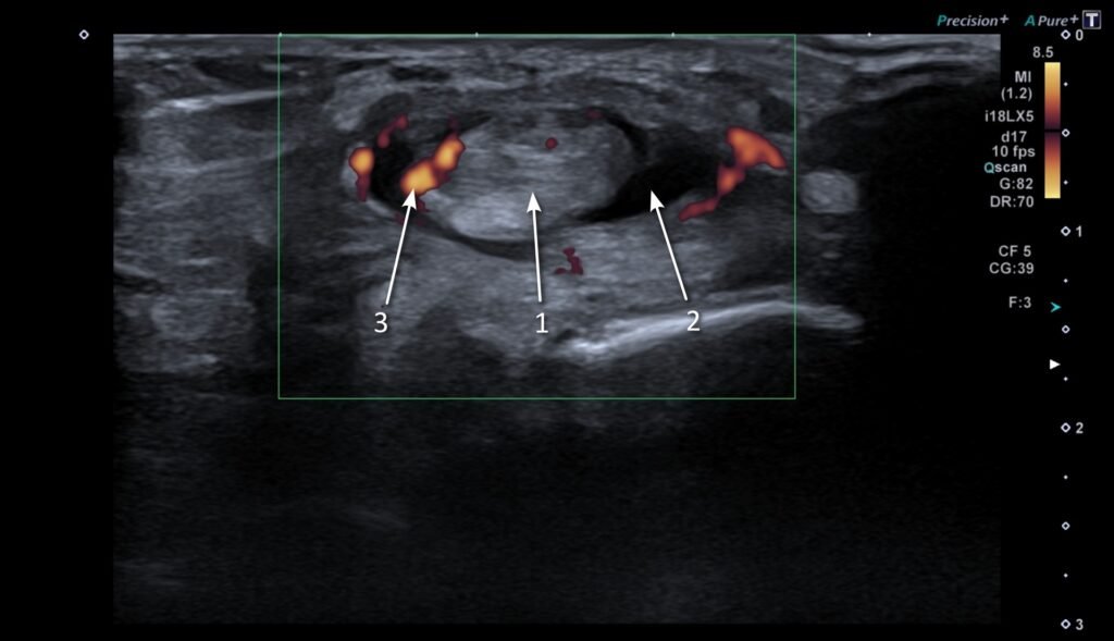
The tendons around the ankle are enveloped by a tendon sheath. This contains a sliver of synovial fluid, a lubricant facilitating the gliding of the tendons during ankle motion. Tenosynovitis is when the tendon sheath itself gets inflamed. On ultrasound, this is seen as a build-up of fluid, thickening and increased blood flow (see Figure 4).
Ligament injuries
Ankle sprains are classified as high and low ankle sprains. In a high sprain, the ligaments connecting the tibia and fibula are affected, the so-called syndesmotic ligaments. In low ankle sprains, the ligaments that connect the tibia and fibular to the talus are affected. The low ankle ligaments are arranged in two groups:
- The medial collateral ligaments, or deltoid ligaments, are on the inner side of the ankle and are injured by eversion-type injuries (the ankle bends outward).
- The lateral collateral ligaments get injured with inversion injuries (when the ankle bends inwards).
The most frequently torn ligament on the outer side is the anterior talo-fibular ligament (ATFL). Failure of proper healing of this ligament can lead to chronic ankle problems, such as chronic pain or instability. Although MRI is the gold standard for assessment of the ankle ligaments, most of these ligaments, and more so the ATFL are well seen on ultrasound. They appear as well-defined, thin bands. A ligament tear is confirmed if there is discontinuity and laxity of the ligament. Figure 5 demonstrates a nice example of a high ankle sprain with a tear of the anterior-inferior tibiofibular ligament (AITFL). Figure 6 demonstrates a low ankle sprain with a tear of the anterior talo-fibular ligament (ATLF).


Plantar fasciitis
The plantar fascia is a strong tendinous band, that extends from the heel bone (calcaneus) to the toes. Plantar fasciitis is a common cause of heel pain. Plantar fasciitis is when there is degeneration and inflammation of the plantar fascia due to overuse. This usually occurs close to its attachment at the heel bone. Ultrasound is as good as MRI in detecting disorders of the plantar fascia. A thickened and dark (hypoechoic) plantar fascia or increased blood flow (Doppler activity) may indicate a potential problem. These changes are often more obvious when the comparison is made to the patient’s other side, which is easily done during the ultrasound examination. Bony spurs at the heel bone are very common but are a factor predisposing to fasciitis. A less common site of fasciitis is where the outer band of the plantar fascia attaches to the base of the fifth metatarsal. This leads to pain at the outer aspect of the foot, very similar to the symptoms caused by tendonitis of the peroneus brevis tendon.
Conclusion
Common conditions around the ankle include Achilles tendon rupture, Achilles tendonitis, tibialis posterior and peroneal tendinopathy, ligament injuries, and plantar fasciitis. Ultrasound can visualise and characterise these issues and perform a dynamic assessment.
Useful links
Achilles tendinopathy | Health topics A to Z | CKS | NICE
Plantar fasciitis | Health topics A to Z | CKS | NICE

Dr M Siebachmeyer
Musculoskeletal radiologist based at St Georges Hospital, London.
References
[1] C. Y. Hung et al., “Advanced Ankle and Foot Sonoanatomy: Imaging Beyond the Basics,” Diagnostics (Basel), vol. 10, no. 3, 2020, doi: 10.3390/DIAGNOSTICS10030160.
[2] A. Aminlari et al., “Diagnosing Achilles Tendon Rupture with Ultrasound in Patients Treated Surgically: A Systematic Review and Meta-Analysis,” J Emerg Med, vol. 61, no. 5, pp. 558–567, Nov. 2021, doi: 10.1016/J.JEMERMED.2021.09.008.